views
Sphingomyelins are the most common sphingolipids in which the terminal hydrocarbon group of the ceramide is replaced by choline phosphate or ethanolamine phosphate. In the sphingomyelin molecule, the amino group of sphingosine forms an amide bond with the fatty acid, and the hydroxyl group of sphingosine is attached to phosphatidylcholine.
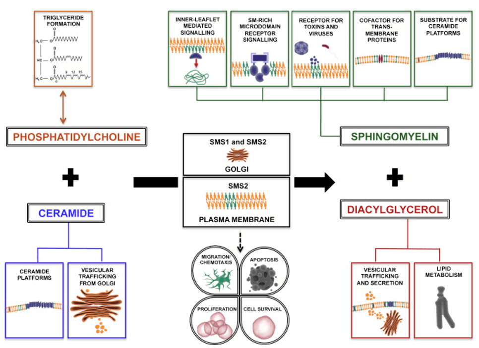
Sphingomyelin is mainly located on cell membranes, lipoproteins and other lipid-rich tissue structures. Sphingomyelin is very important for maintaining cell membrane structure, especially the micro-control function of the cell membrane (such as membrane invagination). It can regulate the activities of growth factor receptors and supercellular matrix proteins, and provide binding sites for some microorganisms, microbial toxins, and viruses. Many extracellular drugs and external stimuli such as tumor necrosis factor (TNFσ), interleukin (IL-1), gamma radiation, interferon (IFNγ), etc. can hydrolyze sphingomyelin and release ceramide by activating sphingomyelinase. Ceramide then continues to perform its physiological functions as a second messenger.
Sphingomyelin is also found in natural foods, and is most abundant in foods such as eggs, meat, fish and milk. Studies have shown that sphingomyelin has a potential inhibitory effect on colon cancer. In addition, sphingomyelin can also reduce serum cholesterol, improve skin barrier function and promote neurodevelopment in infants and young children, making sphingomyelin a potential functional food.
 Cellular functions affected by the sphingomyelin synthase (D'Angelo et al., 2018)
Detection of sphingolipids
High-performance liquid chromatography (HPLC)
HPLC is the most commonly used method for the analysis and detection of sphingolipids, which can be divided into ultraviolet detector (UV), evaporative light scattering detector (ELSD), mass spectrometry and so on according to different detectors.
- HPLC- ELSD
The evaporative light scattering detector is a general-purpose detector capable of analyzing any compound with volatility lower than that of the mobile phase. Its detection does not depend on the optical properties of the sample and is not affected by functional groups, making it suitable for the detection of components with no or weak UV absorption.
- HPLC- UV
UV detector is a detector designed based on the principle that solute molecules absorb UV light, and is used to analyze substances with UV absorption, such as those containing unsaturated structures such as carbonyl, carboxyl, amino, and carbon-carbon double bonds. Phospholipids have UV absorption at 200 nm~210 nm. This method can achieve better separation of SM, phosphatidylcholine, etc, but is influenced by the mobile phase, which is prone to baseline drift, and it requires a certain degree of similarity between the structure of the standard and the sample, which limits the application of the method.
- HPLC- MS
MS uses electric and magnetic fields to separate the moving ions by mass-to-charge ratio and then detects them. This method is mainly used to analyze molecular structures and species of pure substances. The diversity of fatty acid species in the molecular structure of sphingomyelin makes it complicated to perform quantitative analysis alone. In contrast, as an effective separation and analysis method, chromatography enables quantitative analysis of organic compounds, but qualitative analysis is relatively difficult. The combination of the two is more efficient for qualitative and quantitative analysis of complex compounds.
Shotgun method
Direct injection electrospray tandem mass spectrometry is one of the strategies of birdshot lipidomics research, in which samples without chromatographic separation are injected directly into the mass spectrometer through a flow injection pump. The sample is characterized and quantified using the neutral loss scan (NLS) and the parent ion scan (PIS) of the tandem triple quadrupole mass spectrometry. This method is sensitive, fast and without chromatographic separation, and is widely used for lipid analysis.
Sphingomyelin generates product ion fragments of phosphocholine during the collision-activated dissociation (CAD) process. Therefore, the daughter-to-parent scanning mode can be used to determine the corresponding sphingomyelin through the same product ion fragments.
Reference:
- D'Angelo, G., Moorthi, S., & Luberto, C. (2018). Role and function of sphingomyelin biosynthesis in the development of cancer. Advances in cancer research, 140, 61-96.


Comments
0 comment